Read High-Risk Atherosclerotic Plaques: Mechanisms, Imaging, Models, and Therapy - Levon Michael Khachigian | PDF
Related searches:
10 Home Remedies for Plaque and Tartar - Facty Health
High-Risk Atherosclerotic Plaques: Mechanisms, Imaging, Models, and Therapy
Cholesterol and Artery Plaque Buildup
Plaque Psoriasis: Causes and Risk Factors
Prevalence and Characteristics of Carotid Artery High‐Risk
Imaging of high-risk carotid artery plaques: current status and future
Similarity between high-risk atherosclerotic plaque and cancer cells
Vulnerable Plaque, Characteristics, Detection, and Potential - MDPI
MRI in the early identification and classification of high-risk
Risk Factors of Subclinical Atherosclerosis and Plaque - Frontiers
Atherosclerosis Plaque Rupture, Heart Attack and Stroke Risk
Atherosclerosis Plaque Imaging and Characterization Using
Atherosclerosis - Heart and Blood Vessel Disorders - Merck
Coronary CT angiography features of ruptured and high-risk
Atherosclerosis and Stroke: How Excess Plaque May Cause Stroke
Terminology for high-risk and vulnerable coronary artery plaques
Imaging Atherosclerosis and Vulnerable Plaque Journal of
Atherosclerosis and Stroke American Stroke Association
Atherosclerosis Guide: Causes, Symptoms and Treatment Options
Atherosclerotic Plaque Formation and Risk Factors
Lifestyle factors and high-risk atherosclerosis: Pathways and
Carotid plaque imaging and the risk of atherosclerotic
Plaque (fatty deposits) build up in your arteries is called atherosclerosis. These deposits are made up of cholesterol, fatty substances, cellular waste products, calcium and fibrin (a clotting material in the blood).
Despite the small dimension of atherosclerotic lesions, our results suggest that visualization of high-risk plaques in human artery may be possible with α v β 3 integrin-specific spect/ct imaging.
How atherosclerotic plaque causes damage most of the damage occurs when plaques become fragile and rupture. Plaques that rupture cause the formation of blood clots that can block blood flow or break off and travel to another part of the body. In either of these cases, if a clot blocks a blood vessel that feeds the heart, it causes a heart attack.
Dec 20, 2019 5: high-risk prevention: stepped approach or statins therapy down the coronary, and every millimeter of the vessel was atherosclerotic.
Feb 11, 2015 however, they do not affect blood flow for decades, because the artery muscular wall enlarges at the locations of plaque.
Plaque is a sticky, invisible film of bacteria that lives in the mouth and sticks to the teeth. When this slimy coating is left alone, it turns into chalky calcified substance called tartar, which is extremely difficult to remove.
While these play an important role in the progression of atherosclerotic lesions [10, 11], more often, atherothrombosis is asymptomatic and insidious� and can be a warning sign for high-risk plaques which can potentially be detected by umi of activated platelets.
Conclusion: gp iib/iiia of activated platelets on the atherosclerotic endothelium is a biomarker for high-risk plaques that can be quantified by umi using mb-crgds, providing a noninvasive means for detecting high-risk plaques and preventing acute cardiovascular events.
Vulnerable plaques are atherosclerotic plaques that have a high likelihood to cause thrombotic complications, such as myocardial infarction or stroke. Plaques that tend to progress rapidly are also considered to be vulnerable. Besides luminal stenosis, plaque composition and morphology are key determinants of the likelihood that a plaque will cause cardiovascular events.
Developed countries is atherosclerotic cardiovascular disease, commonly caused by thrombotic occlusion of a high-risk coronary plaque, resulting in myocardial infarc-tion or sudden cardiac death, or embolization from a high-risk carotid plaque resulting in stroke.
Nov 4, 2019 hardening of the arteries; arteriosclerosis; plaque buildup – arteries; those with a history of high blood pressure readings or those with risk.
High-risk atherosclerotic plaque features for cardiovascular risk assessment in the prospective multicenter imaging study for evaluation of chest pain trial over the past decade, our understanding of coronary atherosclerosis has evolved from a paradigm dominated by coronary stenosis towards a deeper, more nuanced appreciation of coronary anatomy and physiology.
Atherosclerotic plaques at the highest risk of rupture clearly exhibit a large lipid-rich necrotic core, thin fibrous cap, neovascularization, spotty calcium and abundant inflammatory cells; these.
Given the high risk of sudden death or myocardial infarction associated with atherosclerosis, frequently with no previous symptoms, techniques to better identify high-risk plaques might help lower.
Atherosclerotic lesion, macrophages can release reactive oxygen species and matrix-degrading proteinases. Theseenzymes can remodel the arte rial matrix, facilitate cellularmigration,and likely contribute to thecompensatoryenlargementchar acteristic of the growing plaque.
1 what is an atherosclerotic plaque? a plaque is a regional thickening of the vessel wall caused by atherosclerosis. Plaques are covered by a fibrous cap and consist of ldl-cholesterol, collagen, smooth muscle cells collagen calcium and different cells (monocytes/macrophages, t lymphocytes, neutrophils and foam cells).
Atherosclerotic plaque builds up gradually in the walls of the body's arteries. Ruptured plaque can trigger clots that cause life-threatening conditions such as stroke and heart attacks.
Feb 24, 2016 atherosclerosis; arteriosclerosis; plaque buildup - arteries; hyperlipidemia increased risks include people with high-normal blood pressure.
The lower predictive value of high-risk plaque also reflects temporal changes in cardiovascular disease during the last 2 decades, which includes a shift in acute coronary syndome presentations from myocardial infarctions with st-segment elevation to those without st-segment elevation, decrease in plaque ruptures in culprit lesions of acute coronary syndromes, and more stable characteristics of atherosclerotic plaques (eg, smaller lipid cores and less intraplaque inflammation).
Sep 5, 2017 atherosclerotic plaques: correlation with intra-vascular ultrasound ventional coronary cta high-risk features (low attenuation plaque,.
In contrast to vulnerable plaques in the coronary arteries, high-risk plaques in the magnetic resonance imaging for atherosclerosis plaque characterization.
May 11, 2020 qualitative features of atherosclerotic carotid plaques prone in high-risk plaques were consistent with a change to increased glycolysis.
Atherosclerosis is a general arterial disease and the discovery of plaques in one vascular site should lead to the search for plaques in other sites at risk. Any discovery of a severe atherosclerotic lesion will lead to the search for silent lesions in other locations.
Background carotid atherosclerotic plaque is identified as one of the main sources of ischaemic stroke. However, the prevalence of carotid high-risk atherosclerotic plaque in chinese patients with ischaemic cerebrovascular events has been inconsistently reported and needs to be investigated in a large population. Objectives the primary objective of care ii study was to determine the prevalence.
This is defined as carotid artery disease, which is a form of atherosclerosis. Carotid artery disease is a major risk factor for stroke, because plaque can smoking (past or present); high blood pressure; diabetes; high cholesterol.
Dec 14, 2020 all people aged over 40 years should have a cardiovascular health risk assessment - usually available at your gp surgery.
These plaques may narrow or block an artery, reducing or stopping blood flow. Not all high cholesterol levels increase the risk of atherosclerosis.
Lifestyle risk factors influence plaque composition by modulating traditional and novel pathways associated with cardiovascular risk. Red colour indicates initiation of high-risk atherosclerosis, green colour indicates inhibition of high-risk atherosclerosis.
Atherosclerotic disease, a primary cause of stroke and myocardial infarction, is the most common underlying cause of death worldwide. While atherosclerosis was formerly considered to be a relatively inert structural abnormality, decades of research have since shown that it is a biologically active process, driven by active inflammation.
Atherosclerotic lesions, or atherosclerotic plaques, are separated into two broad categories: stable and unstable (also called vulnerable). The pathobiology of atherosclerotic lesions is very complicated, but generally, stable atherosclerotic plaques, which tend to be asymptomatic, are rich in extracellular matrix and smooth muscle cells�.
Atherosclerosis is a narrowing of the arteries caused by a buildup of plaque. It’s a type of arteriosclerosis, or hardening of the arteries.
Phrases similar to phrases similar to thank you suitable to be printed or engraved on a plaque include in grateful appreciation, with thanks, a world of thanks, your efforts are greatly appreciated, with gratitude and we cannot.
Risk factors for atherosclerosis (eg, dyslipidemia, diabetes, cigarette smoking, in general, unstable coronary artery plaques have a high macrophage content,.
Plaque psoriasis, the most common form of psoriasis, affects about 4 million people in the united states. Plaque psoriasis plaque psoriasis is a chronic autoimmune condition.
Cancer survivors are at increased risk of plaque development and are therefore called high-risk population.
Researchers have developed a tool to identify high risk atherosclerotic plaques, often referred to as inflamed fatty deposits, to help fight cardiovascular disease.
A, kaplan-meier estimates stratified by the presence of high-risk plaque (hrp), adjusted for significant stenosis [ss], defined as 70% or greater stenosis in any coronary artery or 50% or greater stenosis in the left main coronary artery and for atherosclerotic cardiovascular disease risk score.
Jun 22, 2018 of high density lipoprotein cholesterol are associated with subclinical atherosclerosis and plaque burden in high-risk community residents.
It is caused by a buildup of plaque in the inner lining of an artery. You can change some risk factors for atherosclerosis such as smoking, high cholesterol.
Carotid atherosclerotic plaque rupture is an important source of ischemic stroke. However, the prevalence of high‐risk plaque ( hrp) defined as plaques with luminal surface disruption, a lipid‐rich necrotic core occupying 40% of the wall, or intraplaque hemorrhage in chinese population remains unclear.
Carotid atherosclerosis; early-stage atherosclerosis; high-risk plaque; mri; stroke; vulnerable plaque. Stroke is the leading cause of long-term disability as well.
Webmd explains how cholesterol is tied to plaque buildup in the arteries and the medical risks associated with both. Learn how to manage your cholesterol and slow plaque buildup.
Apr 24, 2018 the metabolism of unstable plaque seems to be re-programmed the same way as white blood cells suggesting that treatment with drugs that.
6),7) however, few successes in small-animal imaging of high-risk atherosclerotic plaques have translated clinically. In this review, we will focus on the clinical translatability of molecular imaging of atherosclerosis. First, ideal targets for imaging high-risk atherosclerotic plaques will be briefly discussed.
When a carotid endarterectomy is too risky, doctors may perform a carotid.
Oct 8, 2014 to determine the association between nonalcoholic fatty liver disease (nafld) and the presence of high-risk coronary atherosclerotic plaque.
Mar 7, 2021 atherosclerosis is narrowing of the arteries due to plaque buildup.
The fc is a layer of fibrous connective tissue that separates the lrnc of the atherosclerotic plaque from the arterial lumen. The intact thick fc is associated with a low risk of plaque rupture, whereas a thin fc is associated with a mild risk, and a fissured fc with a high risk of plaque rupture (45,47).
When atherosclerosis completely blocks the brain arteries and/or the above symptoms last longer, it's generally called a stroke. Abdomen — when atherosclerosis narrows the arteries to the intestines, there may be dull or cramping pain in the middle of the abdomen, usually beginning 15 to 30 minutes after a meal. Sudden complete blockage of an intestinal artery often causes severe abdominal pain, sometimes with vomiting, bloody stool and abdominal swelling.
It is not specific to high-risk plaques and is heavily influenced by other factors such as fasting, stress, or medications. Additionally, the myocardium avidly takes up fdg, producing high background signals. Pet imaging of coronary plaques has overcome the motion artifact and partial volume effects due to the small vessel size of coronary arteries.
An abundance of neovessels with elevated expression of integrin αvβ3 is closely associated with an increased risk of plaque rupture. Herein we evaluated the potential of an αvβ3 integrin-targeting radiotracer, (99m)tc-ida-d-[c(rgdfk)]2, for spect/ct imaging of high-risk plaque in murine atherosclerosis models.
Report of a meeting on the vulnerable plaque, june 17 and 18, 2003, santorini, greece. A group of investigators met for two days in santorini, greece, to discuss progress in the field of identification and treatment of high risk/vulnerable atherosclerotic plaques and patients.
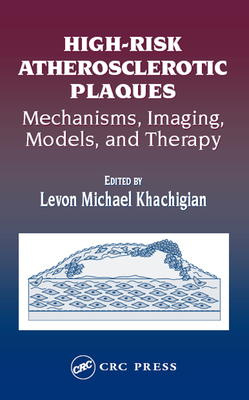
High-risk atherosclerotic plaques: mechanisms, imaging, models, and therapy brings together timely, in-depth reviews by renowned international cardiologists and scientists. Chapters cover the definition, structure, and cellular and molecular mechanisms of high risk plaque development, as well as animal models of vulnerable plaque, plaque imaging, and current and future therapies.
If you're a fan of fried and fatty foods, there's a good chance you have some plaque buildup on the walls of your arteries. If you're wondering how to remove arterial plaque, you may be disappointed with the options.
Feb 16, 2021 the plaque, which is encased by a fibrous cap (collagen smooth muscle cells) is at risk of rupture.
The plaque is mainly composed of fat (or “lipids”) and fibrosis. The risk of plaque is that it may rupture or ulcerate (“unstable plaque”), causing the formation of clots (“thrombus”).
Plaque features predictive of subsequent transient histological studies have shown that many features ischemic attack or stroke during a mean follow-up described in high-risk coronary atherosclerosis are of 3 years (48).
“vulnerable” plaques are atherosclerotic plaques that have a high likelihood to cause thrombotic complications, such as myocardial infarction or stroke. Plaques that tend to progress rapidly are also considered to be vulnerable. Besides luminal stenosis, plaque composition and morphology are key determinants of the likelihood that a plaque will cause cardiovascular events.
Abstract and figures vulnerable plaques are atherosclerotic plaques that have a high likelihood to cause thrombotic complications, such as myocardial infarction or stroke.
High-risk atherosclerotic plaques mechanisms, imaging, models, and therapy 1st edition by levon michael khachigian and publisher crc press. Save up to 80% by choosing the etextbook option for isbn: 9781000611359, 1000611353. The print version of this textbook is isbn: 9780429126413, 0429126417.
High-risk atherosclerotic plaque features for cardiovascular risk assessment in the prospective multicenter imaging study for evaluation of chest pain trial - al’aref - cardiovascular diagnosis and therapy.
The leading cause of major morbidity and mortality in most countries around the world is atherosclerotic cardiovascular disease, most commonly caused by thrombotic occlusion of a high-risk coronary plaque resulting in myocardial infarction or cardiac death, or embolization from a high-risk carotid plaque resulting in stroke.
Feel something weird going on under your hair? we've got answers. These big names didn't let this autoimmune disease get in the way of their success. I feel like the pain is a punishment for my dive into vanity.
Post Your Comments: